|
Cyclic nucleotides (cGMP and cAMP) as intracellular second messengers
All eukaryotic cells utilize cyclic nucleotides—specifically cyclic adenosine monophosphate (cAMP) and cyclic guanosine monophosphate (cGMP)—as intracellular messengers in a wide variety of cell signaling pathways. Learn more... |
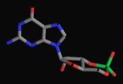
Cyclic GMP
|
Phosphodiesterase (PDE) enzyme superfamily
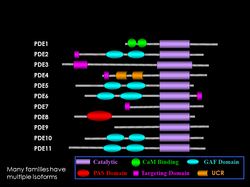
Three major classes of cyclic nucleotide phosphodiesterases (PDEs) have been discovered, Class I, II, and III. The Class I PDEs consist of eleven distinct families found in all vertebrates, and which differ in substrate specificity, tissue localization, regulatory mechanisms, and pharmacological properties. Our lab studies the molecular evolution of PDEs to better understand structure, function, and regulation of this enzyme superfamily. Learn more...
Three major classes of cyclic nucleotide phosphodiesterases (PDEs) have been discovered, Class I, II, and III. The Class I PDEs consist of eleven distinct families found in all vertebrates, and which differ in substrate specificity, tissue localization, regulatory mechanisms, and pharmacological properties. Our lab studies the molecular evolution of PDEs to better understand structure, function, and regulation of this enzyme superfamily. Learn more...
PDE6 is the central effector of the visual signaling pathway in rods and cones
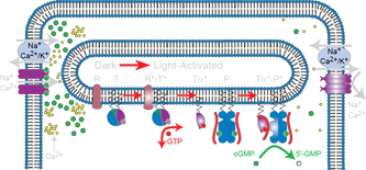
The first step in vertebrate vision is a biochemical cascade of reactions initiated by the absorption of a photon by the visual pigment (opsin) and leading to closure of cGMP-gated ion channels that hyperpolarize the photoreceptor cell and generate a nerve impulse. The photoreceptor phosphodiesterase (PDE6) is subject to precise regulation of its activation (during visual excitation), inactivation (during recovery), and desensitization (during light adaptation) to control cGMP levels in the photoreceptor at each stage of the visual signaling process.
Our lab is integrating biochemical approaches with structural analyses of the macromolecular PDE6 signaling complex in order to define the sequence of steps in the activation, inactivation, and adaptation of the complex of PDE6 and its interacting partners during visual signaling. Learn more...
Defects in PDE6 cause retinal disease but the molecular basis is not understood.
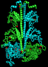
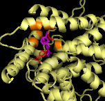
Mutations and adverse environmental factors that impair PDE6 underlie several visual disorders and retinal diseases, such as retinitis pigmentosa and congenital stationary night blindness. Mutations in the catalytic subunits of PDE6 (red spheres in PDE6 structure) have been correlated with disruption of the signaling pathway, often leading to degeneration of the photoreceptor cells and loss of vision. Determining the atomic-level structure of the PDE6 holoenzyme and the topology of the PDE6 signaling complex will aid in the prediction and treatment of retinal diseases and visual disorders that result from dysfunction of PDE6. Learn more...
Mutations and adverse environmental factors that impair PDE6 underlie several visual disorders and retinal diseases, such as retinitis pigmentosa and congenital stationary night blindness. Mutations in the catalytic subunits of PDE6 (red spheres in PDE6 structure) have been correlated with disruption of the signaling pathway, often leading to degeneration of the photoreceptor cells and loss of vision. Determining the atomic-level structure of the PDE6 holoenzyme and the topology of the PDE6 signaling complex will aid in the prediction and treatment of retinal diseases and visual disorders that result from dysfunction of PDE6. Learn more...
Molecular pharmacology of PDEs
PDEs represent excellent targets for pharmacological interventions for several human diseases. A number of family-selective PDE inhibitor compounds targeting specific PDEs have already been approved for clinical use in humans, and many more are in the pipeline. Learn more...
PDEs represent excellent targets for pharmacological interventions for several human diseases. A number of family-selective PDE inhibitor compounds targeting specific PDEs have already been approved for clinical use in humans, and many more are in the pipeline. Learn more...
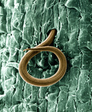
PDEs as novel targets for control of agricultural pests
Although plants use cyclic nucleotides as second messengers in signaling pathways, there are no known Class I PDEs present in any plants for which genomic information is available. This makes PDEs attractive targets for developing compounds that inhibit PDEs present in animal pests that reduce yields of agriculturally important crops. Comparison of the amino acid sequences of nematode and vertebrate PDE families support our hypothesis that compounds targeting plant parasitic nematodes (e.g., root-knot nematode) can be developed that lack adverse effects on vertebrates or on plants. Learn more...
Although plants use cyclic nucleotides as second messengers in signaling pathways, there are no known Class I PDEs present in any plants for which genomic information is available. This makes PDEs attractive targets for developing compounds that inhibit PDEs present in animal pests that reduce yields of agriculturally important crops. Comparison of the amino acid sequences of nematode and vertebrate PDE families support our hypothesis that compounds targeting plant parasitic nematodes (e.g., root-knot nematode) can be developed that lack adverse effects on vertebrates or on plants. Learn more...