A. First steps in vision
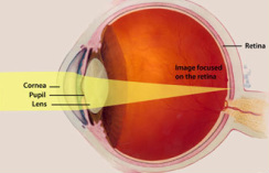
Human vision begins when light enters the eye and is focused by the lens onto the photosensitive tissue at the back of the eye, the retina. Light-sensitive cells in the retina - the rod and cone photoreceptors - capture incoming photons. Through a complex biochemical pathway, the energy of a photon is amplified into a change in the electrical properties of the photoreceptor cell membrane. Changes in neurotransmitter release at the photoreceptor synapse propagate nerve impulses through a series of neurons in the retina. There are five major classes of neurons in the retina: photoreceptor cells, bipolar cells, amacrine cells, and ganglion cells. Eventually, the light signal is transmitted through these retinal neurons to the optic nerve and to the brain.